69 results found with an empty search
- BANGLADESH EMERGENCY CARE SYSTEM IMPROVEMENT (BECSI) PROJECT
Congratulations to A/Prof Gerard O’Reilly, Emergency Physician – Alfred Health, for receiving a project grant from the Australasian College for Emergency Medicine Foundation aimed at promoting emergency care across Bangladesh. Bangladesh has a population of 160 million and is amongst the poorest countries in Asia. It has some of the worst health indices and the burden of injury is substantial. The incidence of disasters is extreme, including cyclones, floods, famine, epidemics, building collapses, bomb blasts and complex emergencies, such as the ongoing Rohingya refugee crisis. There is no effective emergency care system, so there is minimal resilience to these disasters. The ‘Bangladesh Emergency Care System Improvement’ project (BECSI) will be carried out in collaboration with the World Health Organization (WHO) and key emergency care stakeholders in Bangladesh. The stakeholders in Bangladesh remain desperate to improve emergency care in Bangladesh, starting with: A national consensus on current priority actions for emergency care system improvement The introduction of an emergency care system improvement program The BECSI project will use the WHO Emergency Care System Assessment (ECSA) process, which has been conducted in over 30 countries, yielding feasible priority actions and practical next steps for emergency care system development. The BECSI project will promote the development of emergency care in Bangladesh by: Providing a national forum for multi-sector (government, policy, hospital, management, disaster response, prehospital, clinicians) engagement, consensus and an agreed report for emergency care system improvement, and Supporting national representation and participation in the introduction of an Emergency Care System Quality Improvement program. This 12-month project will commence in April 2020. Learn more about this project
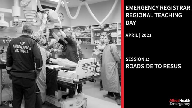
- REPLAY THE EMERGENCY REGISTRAR REGIONAL TEACHING DAY: APRIL 21 | 2021
The Alfred Emergency & Trauma Centre is please to offer the opportunity to replay the online delivery of our recent Emergency Registrar Regional Training Day. This half day program was broken up into the three sessions outlined below: SESSION 1 – ROADSIDE TO RESUS Case based pause and discuss simulation involving the management of a shock-trauma patient with pelvic trauma and retroperitoneal bleeding. Our panelists include Dr Joseph Mathews, Dr Fatima Rahman, Dr Mike Noonan, Dr Lexi Nikolsky, Dr Divya Karna and Ben Meadley. The session is facilitated by Dr Shane Broderick. Recorded on April 21, 2021 SESSION 2: IMAGING, INTERVENTIONAL RADIOLOGY AND SURGICAL MANAGEMENT OF PELVIC FRACTURES Session two of our regional registrar teaching features some radiology pearls from IR fellow Matt Lukies, followed by a panel discussion on the management options of pelvic injuries and a urethral injury. Our panelists include Dr Joseph Mathews, Dr Fatima Rahman and Dr Laura Scott, The session is Facilitated by Dr David McCreary. Recorded on April 21, 2021 SESSION 3: LIGHTNING LEARNING Session three comprises a round of lighting learning with the following topics: C-Spine Clearance – Dr Hector Thomson Approach to junctional bleeds – Dr Danny Marhaba Approach to the managent of suspected airway burns – Dr Jacqueline Morel Procedure – lateral canthotomy – Dr Grace Wong Our panelists include Dr Eleanor Junckerstorf, Dr Vanessa Phua, Dr Andreas Tscharke, Dr Marc Schnekenburger and Dr Shane Broderick. The session is Facilitated by Dr Luke Phillips. Recorded on April 21, 2021
- NECK RADIOGRAPHS: THE STRIDULOUS CHILD
Dr Elizabeth Sheffield Senior Registrar Peer Reviewer: Dr David McCreary CASE OF THE DAY: It’s 23:00, when a 3-year old child is brought in by ambulance with a 2-hour history of noisy breathing, fever and cough. On initial assessment she has inspiratory stridor, is miserable and sitting with her head in a sniffing position, without obvious drooling. She’s previously fit and well, and parents tells you that immunisations are up-to-date. You glance at her vitals and note she is febrile at 38.6, heart rate 180, respiratory rate 38 and oxygen saturations 98% room air. You prescribe your tried and tested cocktail of 0.6mg/kg dexamethasone and 5mls of 1:1000 adrenaline neb then stand triumphant at the foot of the bed basking in self-congratulation…unfortunately 15 minutes later the child is not running around the department in the throws of a steroid-induced mania and remains cuddled up to mum/dad displaying much the same picture as before. WHAT IS THE HIGHEST PRIORITY DIAGNOSIS? You can easily take your pick – but this is a toxic child. Sniffing position triggers an exam answer of retropharyngeal abscess – however epiglottitis and inhaled foreign body could certainly give this picture in the right circumstances. Although a bit young for the average bacterial tracheitis it’s definitely within the realm of possibilities. You decide there is still ambiguity with regards to this patient’s diagnosis, and are looking for a handy way to screen for potential sinister causes that are quick, fast and non-distressing to the child. So, behold the soft tissue x-rays. This is a historical Part 2 Exam favourite cropping up every few years in some form or another. Just to signpost, for what conditions might soft tissue neck x-rays be used with regards to acute diagnoses in the Emergency Department? Epiglottis Retropharyngeal abscess Tracheobronchial or oesophageal foreign bodies Bacterial tracheitis Croup (to screen for alternative causes) Neck tumours (although not usually emergent) SO… WHAT MAKES KIDS AT RISK? They lack all sense of self-preservation They have some difficulty differentiating between “food” and “not food” They LOVE to move. They pick-up everything and put in in the nearest orifice, which is (occasionally) their mouths… They may be nonverbal, or at the very least unable to adequately communicate to caregivers that they have choked on a They’re not just tiny adults: their trachea has a smaller diameter, with immature pliable tracheal rings which make them more prone to hyperflexion/extension and more at risk of airway occlusion from any inflammatory pathology. The epiglottis is large and floppy making direct intubation methods (straight blade) different, and also makes them more likely to choke on their own soft tissue… and their larynx is higher and more anterior (and smaller) so surgical cricothyroidotomy is out til about age 10. LET’S TALK GENERAL AIRWAY ANATOMY. Below is a refresher on anatomy of the head and neck... (Courstesy of: https://www.slideteam.net/0514-lateral-cross-sectional-view-of-head-and-neck-laryngeal-anatomy-medical-images-for-powerpoint.html) And here we have the structures as seen on a normal neck x-ray: (Courtesy of https://radiologykey.com/imaging-soft-tissues-of-the-neck/) SO WE’VE GOT THE X-RAY, NOW WHAT? Let’s discuss some handy tips to approaching that neck soft tissue radiograph: SOME ADDED NOTES… Ensuring that breath has been held in inspiration is key to avoid false positives Loss of lordosis or pseudosubluxation indicates the patient is holding their neck in such a way as to avoid collapsibility of their boggy oedematous airways Always pay special attention the epiglottis and the pre-epiglottic space (valleculae). The epiglottis should be a thin structure and there should be a nice space just adjacent to it which is the valleculae. Epiglottitis causes those terrifying laryngoscopic images, and this oedematous soft tissue causes a large “thumbprint sign” (a.k.a. boggy oedematous mass) where the epiglottis should be, as well as narrowing of that nice clean valleculae space. WHEN MIGHT WE GO FOR SOFT TISSUE X-RAY OF THE NECK OVER CT? Well, unfortunately, it’s that classic medical answer: “it depends”. If the child is drooling and holding themselves in a tripod position, then you aren’t going to want to mess around with a CT – it’s going to be straight to the operating theatre for a gaseous induction with anaesthetics and ENT on stand-by… in the ideal world. With regards to imaging in the more stable patient, my personal experience has been X-ray first, and if there is ambiguity – and the child can tolerate it – then CT is the next line... Broadly speaking, X-ray has some advantages when: you want to minimise ionising radiation the patient is too scared to go to the scanner any airway apprehension meaning the patient cannot lie flat you want a quick test you don’t have CT available SIDE NOTE: RETROPHARYNGEAL ABSCESSES – FROM A SYSTEMATIC REVIEW CO-AUTHORED FROM OUR OWN RAMANAN DANIEL (OTOLARYNGOLOGY). A systemic review (1) comparing X-ray and CT to gold standard of intra-operative pus found high sensitivity and specificity rates in several studies (ranging from 80 to 100% (for all but one study)). The exception to the above was a single study performed by (Ravindranath et al 1993 (3)), was very much an outlier, finding a sensitivity and specificity of 0%. A question mark may be able to placed next to their study results however due to small sample size of only 10 patients. The authors the systematic review have stated that potentially those finding a higher sensitivity and specificity for X-ray had populations that tended to be severely unwell (however there was not sufficient clinical data in any paper to firmly make this conclusion). Essentially, if an effort has been made to obtain as high a quality radiography as possible (with appropriate rotation, neck extension, and respiratory phase) then X-ray in the Emergency Department can prove to be extremely useful in identifying patients requiring theatre for drainage of RPA. NOW, THE FUN STUFF. LET’S LOOK AT A FEW EXAMPLES: (Case courtesy of Dr Maxime St-Amant, Radiopaedia.org, rID: 26840) CAN YOU LABEL THE RELEVANT ANATOMICAL LANDMARKS ON THE ABOVE IMAGE? This patient has epiglottitis. NB there is also loss of normal cervical lordosis. Along with above check the subglottic airway to check for oedema and narrowing of the subglottic space inferiorly. (Case courtesy of RMH Core Conditions, Radiopaedia.org, rID: 26246) WHAT PATHOLOGY DO YOU SEE ON THE ABOVE RADIOGRAPH? This is a subtle retropharyngeal abscess. Again there is loss of normal cervical lordosis, although C1 and C2 look reasonably normal. Differential diagnoses: any cause of fluid, so trauma, hereditary angioedema, anaphylaxis or neoplastic disease in that retropharyngeal space. (Case courtesy of Dr Ian Bickle, Radiopaedia.org, rID: 30018) WHAT ARE THE MAIN ABNORMALITIES ARE ON THE ABOVE IMAGE? This is a retropharyngeal abscess (Case courtesy of Assoc Prof Frank Gaillard, Radiopaedia.org, rID: 6258) WHAT FINDINGS ARE THERE ON THE ABOVE IMAGE? Distended hypopharynx and subglottic narrowing. There is no swelling of the prevertebral space, the epiglottis is very normal looking and there is a nice and full valeculae. Differential diagnoses will include viral croup or bacterial tracheitis depending on clinical picture. (Case courtesy of Dr Michael Sargent, Radiopaedia.org, rID: 6086) AND FINALLY, WHAT DO YOU MAKE OF THIS ONE? Another croup X-ray, this time an AP. Some stenosis is normal, so don’t get caught out when you see it when not clinically indicated, but should not be pronounced. Also, if you see similar in a slightly older child but with a toxic presentation, don’t forget about that ugly stepsister of the croupy child, bacterial tracheitis. SO – YOU ACED IT. BUT WHAT ARE WE GOING TO KNOW WITH OUR NEW KNOWLEDGE… ARE WE GOING TO ORDER A SOFT TISSUE X-RAY ON THAT NEXT CHILD WE SUSPECT HAS CROUP? Well, is there a prodrome of coryza, fever and progression to barking cough in a non-toxic child who attended daycare a few days ago? If so, you can likely rest assured with your clinical diagnosis of croup. If however, you have a toddler who developed a barking cough after playing unsupervised in the garden, maybe with a stick in his hand 2 days ago, no viral illness or clear precipitant, then the lateral soft-tissue is a pretty reasonable go-to as an initial screening test to aid future management… that, and perhaps a baby leash. CLINICAL PEARLS Sometimes you need to have a sneaky plan with radiology and ENT and just wait for the kiddo to have a nap and then do the CT when they are asleep Always discuss with ENT early if they can come in and consider performing a naso-endoscopy in the child who you feel may be appropriate (read: not going to obstruct their airway in ED) Don’t forget button batteries. Ingestion can present in the vaguest of ways, even just being “off food”. Have a high index of suspicion and have a low threshold to observe or obtain images on the kid who really doesn’t quite fit or seem right. SOME FOOD FOR THOUGHT How might you approach the patient who requires sedation for transport or imaging? What agent would you use and how might you manage this in a resource limited setting (no anaesthetist, no ENT)? What would be your approach to the emergent and also crash airway? What options would you consider? FUTURE SCOPE: USS IN THE ED? Limited data on the utility of this… but a developing space. Experiences detailed in a case series in the American Journal of Emergency Medicine (2) suggest that POCUS may aid and expedite medical decision making in the undifferentiated patient. Specifically, the technique (as you would assume) involved identifying a hypoechoic collection or hypoechoic/anechoic collection in the prevertebral space. At this stage, cannot be recommended as an alternative to other modalities of imaging but an interesting area to follow… Fig 3: A&B hypoechoic collection with irregular borders | Fig 4: A&B circular heterogenous collection with effaced IJ | Right hand image: transducer placement for evaluation of RPA (https://www.ajemjournal.com/article/S0735-6757(20)30657-4/fulltext) KEY REFERENCES: 1) The accuracy of lateral X-Ray and computed tomography in diagnosis of paediatric retropharyngeal abscess: a systematic review 2) A novel approach: Point-of-care ultrasound for the diagnosis of retropharyngeal abscess 3) Computed Tomography in Diagnosing Retropharyngeal Abscess in Children ELIZABETH SHEFFIELD Senior Registrar, Alfred Health Dr. Elizabeth Sheffield (BSc (Hons), MA, MA (Hons), MBChB) is a critical care and airway enthusiast from Washington, D.C., working at The Alfred Hospital in Melbourne, Australia. Lover of medical education, SIM, rural medicine and critically ill airway management in the ED, plus exploring the outdoors with her beautiful family and frequently wet dog.
- IV FLUID ADMINISTRATION: PAEDIATRIC & NEONATAL
Dr. Elizabeth Sheffield Senior Registrar PEER REVIEW: Dr Emma Bellenger Editor: DR DAVID MCCREARY CASE OF THE DAY: You’re on night shift and your short stay resident is concerned that their 3 year-old patient has pyelonephritis. They have been vomiting continuously despite 4 mg of ondansetron. Bedside urine confirms leukocytes, nitrates, blood and ketones. On exam, the child is alert but miserable, flushed, with dry oral mucosa. They have a RR 30, SBP 100, HR 150, & CRT 2 seconds centrally. On your assessment, the child is unwell, but not shocked, so you ask the resident to commence IV benzylpenicillin 60 mg/kg and IV gentamicin 7.5 mg/kg and IV fluids. The resident hasn’t had much paediatric experience and asks you to instruct them how best to tackle ongoing rehydration. WHY DOES IT MATTER? I.V. fluid is a drug (ie. it has associated benefits, side effects, indications, contraindications and complications). It’s true what they say: kids have brains like a sponge… excessive or incorrect fluid administration (and subsequent hyponatramia) can lead to cerebral oedema, seizures and death. It is an evolving area of medical research and as such there can be a lack of consistency in the guidelines across states, hospitals and sometimes departments. The ACEM Part 2 exam is typically riddled with paediatric fluid calculation MCQs… which is why it’s good to nail down a quick strategy to answering questions that won’t cause you too much of a headache during the exam when you’re already under pressure. OUR STRATEGY FOR FLUID REPLACEMENT CAN BE SUMMARISED AS FOLLOWS: Treat shock. Replace deficit in addition to ongoing maintenance fluids over 24 to 48 hours. Account for ongoing losses. 🤓 We’re focusing on IV hydration in this post, but remember enteral fluids via NGT are often preferred. For more on NG fluids check out the RCH guideline here. WHICH PATIENT? Generally, IV fluids are required for patients who: are NBM (e.g. pre-surgery) are failing an oral fluid challenge and won’t will not tolerate an NG tube have significant dehydration or shock which needs urgent correction WHICH CONDITIONS SHOULD BE EXCLUDED FROM THE THE STANDARD FLUID CALCULATIONS? Diabetic Ketoacidosis Burns Haemorrhage or significant trauma Known or suspected inborn errors of metabolism Renal failure Liver failure Pyloric stenosis Oncology Hyperhydration Premature children < 1 week corrected age These conditions require specialist advice and often have their own guidelines or will involve discussion with a paediatrician. WHICH FLUID? ISOTONIC VS HYPOTONIC FLUIDS? Avoid Hypotonic fluids such as 0.45% sodium chloride or straight 5-10% glucose without additives. Prescribe isotonic fluids such 0.9% sodium chloride, Hartmann’s or Plasma-lyte 148 – to which we typically add 5-10% glucose. ISOTONIC FLUIDS – WHAT’S THE DIFFERENCE AND WHY DO WE CARE? It’s all about balance. Let’s compare our plasma to what’s going on in these fluids… The main difference you should note is our serum chloride of 103 mmol/l vs the 154 mmol/l of chloride you’re getting with 0.9% saline. Excess chloride can cause subsequent development of hyperchloraemic metabolic acidosis, which may reduce renal perfusion and cause acute kidney injury. The studies in this area are not robust enough to be conclusive at this stage. With evidence as it sits now, all 3 are acceptable fluids to administer and in ED we’re unlikely to be keeping patients long enough to give large volumes of fluid compared to in intensive care or on the paediatric ward, so our initial choice of fluid therapy has so far not been weighted to one or the other. WHICH FLUID DO WE CHOOSE FOR DEFICIT REPLACEMENT AND MAINTENANCE THERAPY? In contrast to our adult patients, with these kids we always need some glucose in the mix. The question as to whether that is 5% or 10% (or more) is a bit murky depending on how young the patient is. For exam purposes: For children < 4 weeks corrected age: the fluid of choice is an isotonic (e.g. 0.9% chloride) + 10% glucose, +/-20 mmol/L potassium chloride. For children > 4 weeks corrected age: the suggested choice is 0.9% + 5% glucose (although Plasmalyte-148 + 5% glucose, or Hartmann’s + 5% glucose are accepted alternatives in some institutions and interchanging these can be a trick on exams). WHAT’S WITH THE SUGAR? Infants and children have a higher consumption of glucose per kilogram than adults. Neonates have even higher requirements still and are at greater risk of becoming hypoglycaemic, hence the requirement for 10% glucose. DO WE ADD POTASSIUM? In the ED, generally not, unless the patient is hypokalaemic on their UEC. WHICH RATE? You need some calculations for the total base amount of fluid to be given: Bolus (if required): 10-20ml/kg x 2-3 doses Deficit (if required): weight (kg) x % deficit x 10 Maintenance: 4:2:1 rule (see below) Any ongoing losses ASSESSING DEFICIT MAINTENANCE: THE 4:2:1 RULE This is a weight-based calculation for determining the general IV fluid maintenance requirement for a paediatric patient per hour. For example - if we take our 3 year old, 14 kg patient above: First 10 kg at 4 mls/kg/hr = 40 mls + Next 4 kg at 2 mls/kg/hr = 8 mls … So, the total IV fluid maintenance rate is 48 mls/hr WHAT ABOUT THE NEWBORNS? In the first week of life, use an algorithmic rather than weight-based guide to maintenance fluid requirements: Day 1: 60 ml/kg/day Day 2: 90 ml/kg/day Day 3: 120 ml/kg/day Day 4-7: 150 ml/kg/day ☝️ Note these are per day so divide by 24 for the hourly rate. After the first week (adjusted) you can use the weight-based 4:2:1 rule to guide your starting doses. Keep in mind there isn’t a one-sized-fits all formula for human fluid requirements. Some patients may have requirements more or less than this to maintain desired clinical end-points. WHEN DO WE USE 2⁄3 MAINTENANCE? Antidiuretic hormone (ADH) is frequently released in our unwell kids – in particular those with meningitis or pneumonia. This release of ADH means that full maintenance is often unnecessary and thus we should consider starting the maintenance fluid aspect of our calculations at a reduced (2/3rd of total calculated based on 4:2:1 rule) dose, and reassess and increase as required. WHO NEEDS EVEN LESS – ½ MAINTENANCE? 50% maintenance should be considered for: patients with suspected meningitis, encephalitis or head injury intubated and cannot tolerate enteral feeding hyponatraemia, discuss these patients with specialist team (PICU, endocrine) in whom hyponatraemia or respiratory distress worsens despite on 70% rate children with inborn errors of metabolism children on dialysis or post renal transplant (reference 1) LET’S RECAP: THIRSTY FOR MORE? We can’t really discuss IV fluids for kids without a discussion of the FEAST trial. This RCT conducted in Africa examined boluses of saline vs albumin vs no bolus. They concluded that there was increased mortality in patients receiving an IV bolus. This was in a population of children with a high incidence of Malaria and severe anaemia, however, and probably isn’t directly applicable to an Australian paediatric population. 🤓 THERE’S A BIT MORE TO IT THAN JUST ‘FLUIDS ARE BAD’, HOWEVER, SO DROP-DOWN FOR MORE INFORMATION IF YOU’RE INTERESTED DESIGN: RCT P: Children 60d – 12y with (all three of): severe febrile illness impaired consciousness and/or respiratory distress impaired perfusion Key exclusions: gastroenteritis, non-infectious causes I: Two groups: Without severe hypotension: 20ml/kg normal saline or 5% albumin With severe hypotension: 40ml/kg normal saline or 5% albumin Severe hypotension = BP<50 (<12 months), <60 (1-5 yrs), <70 (>5 yrs) 🤨 Halfway protocol change alert! Increased to 40ml/kg and 60ml/kg when preliminary results were showing no difference C: No bolus O: Mortality at 48 hours RESULTS: 48 hr mortality significantly worse in bolus groups vs control: 6% (Albumin) vs 10.5% (Saline) vs 7.3% control RR of death saline vs control: 1.44 <95%ci 1.09-1.9,="1.09-1.9," p="0.01"> AUTHORS' CONCLUSION: In the resource-limited setting, fluid bolus significantly increased mortality of children with severe febrile illness and impaired perfusion. 🤓 Editor’s Thoughts: This study has a couple of weaknesses preventing generalisability or application to our paediatric population and it certainly hasn’t changed my practice: As we mention above there was a high incidence of malaria and severe anaemia in this population. Fluid boluses without transfusion may cause more haemodilution in already anaemic patients. The protocol change midway is concerning. Particularly when you see the volume of boluses being tested which have then gone to show increased mortality. I don’t think any of us would be using 60ml/kg boluses in our usual practice, even for severe hypotension. The trial was performed in a resource limited setting with no access to PICUs. SOME CLINICAL PRACTICE POINTS: Don’t forget to factor in the fluid given with medications (antibiotics etc.). Adjust maintenance rate accordingly. Fluid balance should be totalled at least every 6-12 hours, this includes in the ED, particularly in times of access block. Unwell children should have electrolytes and glucose measured 4-6 hours after commencement of IV fluids. Consider whether your patient needs full, 2⁄3 or ½ maintenance Serial weights are the best measure of acute changes in fluid status. Weights should be “bare weight” on babies. Beware sodium. If it’s <135, >145 or changes >0.5 – escalate to your senior or a specialist. IF YOU HAVE THE TIME AND WANT TO TEST YOURSELF, TRY OUT THESE EXAMPLES: Example One: A 2 week-old child born 40+3 is being admitted for suspected meningitis. He is lethargic, and rejecting breast and bottle feeding. His SBP is 70, HR 160 and RR 40, Cap refil is < 2 seconds, and he is peripherally warm. His fonatelle and eyes are sunken. Review of the Maternal Child Health record states a recent pre-morbid weight of 5000g. Bare weight in the department today is 4750g Shocked or not? Nope, no fluid bolus required. What is the deficit? In teeny chippers (newborn) bare weights can be relied upon to determine fluid deficit accurately. Based on the above weights, we have a deficit of 5% (which correlates clinically). Thus, deficit = (pre-morbid weight – current weight)kg x 1000 mls = (5 – 4.75) x 1000 = 250mls If replacing fluid over 48 hours: Thus, rate of replacement of existing deficit per hour would be: = 250mls/48 = 2mls/hour What is the maintenance? Using the 4:2:1 rule: 20mls/hour. Adjusting to 2/3 maintenance = 13.3mls/hour Rate of admin to account for maintenance fluid plus replacement of deficit over 48 hours would be: 13.3 + 5.2 = 5mls/hour. Ongoing losses? Record losses over the space of an hour and then account for those losses over the next hour. If child vomits once in one hour, we would presume a 5ml loss and replace this over the next hour (adjusting the rate to 23.5mls/hour for one hour before reverting back to 18.5mls.hour) Example Two: A 7 year-old child is being admitted with suspected appendicitis. She is haemodynamically stable, with nil concern of shock or perforation. Initially assessment did not reveal any dehydration. Due to access block, she has remained in your department for the past several hours. Maintenance fluid were started 4 hours ago and during this time she has vomited 2 cups of fluid totalling 100mls. She weighs 20kg. Shocked or not? No Deficit? None What is the maintenance rate? Calculated according to 4:2:1 rule is 60mls/hour How does the rate change given the ongoing losses? Given the loss of 100mls over the past 4 hours, we would increase the rate to replace this over the next 4 hours (ie by 25mls/ hour) Thus rate for the next 4 hours will be 60 mls/hour of 5% dextrose saline plus an additional infusion of normal saline at 25mls/hour. Example Three (our 14kg child from the case): Back to our 3 year-old, 14 kg child above. An hour or so later (having been unattended during this time) the nurses alert you she is now in met call criteria with RR of 42, HR 175, and a SBP of 65. Shocked or not? Yes, fluid bolus of 20mls/kg indicated. Second bolus can be considered if shock persists. What is the remaining deficit? Following treatment and correction of shock (which is a > 10% deficit), it can be assumed that the child now has moderate dehydration, which estimated to be 5% (or 50mls/kg) Thus deficit = 14 x 50mls= 700mls Replacing deficit over 48 hours provides a rate of 14.5mls/hour What is the maintenance rate? Using 4:2:1 rule: 48mls/hour What is the total ongoing rate? 5mls/hour over the next 48 hours, whilst monitoring and adjusting for ongoing losses. (whilst monitoring and accounting for ongoing losses) GREAT JOB, YOU ACED IT! RESOURCES: The Starships Hospital guidelines on I.V. fluid administration The Nice guidelines on I.V. fluid therapy The RCH guidelines of I.V. fluid therapy The Queensland Children's Hospital guidelines on I.V. fluid therapy Don’t Forget the Bubbles summary on the evolution in practice regarding I.V. fluid administration in Children. The FEAST Trial - Maitland K, Kiguli S, Opoka RO, Engoru C, Olupot-Olupot P, Akech SO, et al. Mortality after Fluid Bolus in African Children with Severe Infection. New England Journal of Medicine. 2011 Jun 30;364(26):2483–95. DOI: 10.1056/NEJMoa1101549 The Bottom Line review of the FEAST trial ELIZABETH SHEFFIELD Senior Registrar, Alfred Health Dr. Elizabeth Sheffield (BSc (Hons), MA, MA (Hons), MBChB) is a critical care and airway enthusiast from Washington, D.C., working at The Alfred Hospital in Melbourne, Australia. Lover of medical education, SIM, rural medicine and critically ill airway management in the ED, plus exploring the outdoors with her beautiful family and frequently wet dog.
- WILL THAT SCAN GIVE ME CANCER?
DELVING INTO THE RADIATION EXPOSURE FROM COMMON ED IMAGING MODALITIES Dr Emma Bellenger Emergency Registrar Peer Reviewer: DR David McCreary AS IS TRADITION, A POEM TO GET US IN THE MOOD. We send our patients off to scan ‘cause that’s what we do as part of our plan But what is the risk of doing a ct, chest x-ray or angiography? There’s radiation exposure that damages DNA, and may cause cells to mutate in a carcinogenic way But how exactly do we convey this risk of cancer to our patients in a meaningful way Well, you have more of a chance of getting hit by a car but with accidental drowning the risk is on par That’s not to say scans are all bad they help us diagnose for which we are glad So stop and think scans are not risk free weigh up the risk vs harm and use them cautiously. By Emma Bellenger (The ED poet) Every day I send patients for X-rays or CT scans to help with the diagnosis of various conditions. I often mention the “risk of radiation” to my patients, but truth be told is I’ve never really known how much risk there actually is. I’ve managed to elude needing to know this for 5 years now, but recently I was caught out when a patient asked me, “so how much risk are we talking, doc?” At which point I blushed, shrugged and told them “pretty low risk” before slowing backing out of the room. So if you, like me, are ready to add some facts to that “at risk” spiel, then this is the blog for you. I have aimed to not get blogged down in the numbers but instead represent the risk in a way that is relatable and meaningful to us all. But first.. WHY IS IONISING RADIATION HARMFUL? 🚨 Trigger warning – primary exam knowledge 🚨 Ionising radiation → ionised atoms → free radicals → DNA damage → mutation → cancer As the name suggests, ionizing radiation causes an atom to become ionised (unstable - missing an electron). This can lead to the production of free radicals and subsequent tissue damage through reactions with DNA. Dividing cells are most vulnerable to these processes and can result in cell death or mutation that can then lead to carcinogenesis. Picture source: https://crossfittiltiii.com/news-events/coach-kathleen-on-antioxidants/ WHAT FORMS OF IMAGING USE IONISING RADIATION? Xray and CT are our main culprits within the ED. Angiography and fluoroscopy, too – but that’s not something we use frequently within the department. Relevant negatives are USS and MRI which are both free of ionising radiation. HOW MUCH RADIATION? In day-to-day life we are all exposed to a degree of background radiation. The amount is dependent on many different factors, but the average Australian is exposed to 1.5-2 mSv per annum. (source: arpansa.gov.au) One potential way to explain to patients how much radiation a scan will expose them to, is by relating the amount of radiation to a year’s supply of background radiation. For example, a CT chest is equivalent to about 4 additional years of background radiation. If you feel this would work for your spiel, the table below from the American College of Radiologists represents these figures nicely: There are a couple of other tables from NSW health or the FDA you can check out if this one isn't to your liking. C’MON, GIVE THE PEOPLE WHAT THEY WANT, WHAT’S THE RISK OF ALL THIS RADIATION? So now that we know how much radiation certain scans add to our background radiation exposure, we need to know how we can convey this in a meaningful way in terms of risk. And what we really mean by that is, what’s the risk of getting cancer?! Well, turns out radiation is not as carcinogenic as many people think (or at least as I thought). The risk depends on many factors including the person’s age, gender and part of the body being exposed; however, most sources agree that a CT will increase a person’s risk of fatal cancer to between 1 in 1000 to 1 in 2000 people. When you compare this to the life-time risk of getting cancer anyway, 400 in 2000 people (or 1 in 5), this is a low risk (FDA.gov). For those of you who are still sceptical about the risk, how many of you have ever thought you would die from accidental drowning? I’m guessing not many…and turns out that’s approximately the same likelihood as dying from cancer from imaging radiation exposure. Source: https://www.racgp.org.au/afp/2013/june/radiation-safety/ WHAT ABOUT HIGH-RISK POPULATIONS? Pregnancy, infants and children make up a group where exposure to radiation is a particularly worrisome thought. The figure below shows that young girls are of highest risk due to the nature of dividing cells, in particular breast tissue (1 in 100 risk of cancer from 1 CT scan). Comparatively, elderly people have extremely low risk of cancer, largely due to remaining life expectancy limiting their chances of developing cancer before they die. Source: https://www.racgp.org.au/afp/2013/june/radiation-safety/ The concern with pregnant ladies is the harm caused to the foetus. Detrimental tissue effects from foetal exposure to radiation have only occurred at doses greater than 100msv. All imaging in pregnancy should be discussed with a senior clinician and may need input from your radiologist ± an obstetrician, but the graph and table below gives us a brief overview of the stats. Source: RANZCR, Inside Radiology: Radiation Risk of Medical Imaging During Pregnancy CTPA VS VQ SCAN FOR INVESTIGATING PE IN PREGNANCY? It's worth being aware of this for a couple of reasons: It's a common clinical conundrum It's a common exam conundrum Let's look at the estimated radiation doses and risk from CTPA vs VQ scan in pregnancy: Source: RANZCR - Suspected Pulmonary Embolism Given that the foetal radiation exposure is minimal in both scans, the decision comes down to maternal risk. For this reason, VQ scan is recommended over CTPA as it limits radiation to the breast tissue. SUMMARY Imaging in the emergency department is an important part of our workup for many patients. As we have learned, the risk of cancer is relatively low, none the less it exists and thus scans should only be ordered when they are justified. There are plenty of ways to express the radiation exposure to our patients – be it through comparing the radiation to yearly background radiation, chest X-ray equivalents (see table below), or through comparing the risk to other ways of dying (morbid, right?). SUMMARY OF RADIATION EXPOSURE OF COMMON ED IMAGING MODALITIES EXPRESSED IN CHEST X-RAY EQUIVALENTS So next time a patient asks you what is their risk of getting cancer from a scan, no more backing out of the room slowly… you can hit them with the facts; ask them how often they go swimming; and help them make an informed decision. Picture source: https://ctscanmachines.blogspot.com/2018/06/ct-scan-funny.html EMMA BELLENGER Emergency Registrar, Alfred Health Dr Emma Bellenger is an Emergency Registrar at the Alfred Hospital with a passion for life long learning, quality of life and efficiency. Alongside her role in ED she dabbles in sports medicine working with various football teams. Pre-pandemic Emma enjoyed doing hikes around the world, but of recent times has found solace in the simple things in life such as trail running, cuddling her cats and LEGO.
- ALTITUDE SICKNESS
Dr Danny Marhaba Emergency Registrar Peer Reviewer: Dr David McCreary Weekly Advanced Group teaching at the Alfred includes a lightning learning presentation from one of our registrars. From time to time, we’ll ask them to record these talks to share their teaching with the FOAMed world. This week featured a great talk from Dr Danny Marhaba on Altitude Sickness. Albeit not a presentation we will see often in Australia, but an important one nevertheless - particularly if you are expedition-medicine-inclined. IN SUMMARY: DEFINITIONS: HAH: High Altitude Headache (An isolated headache due to hypoxic cerebral vasodilation). AMS: Acute Mountain Sickness (HAH + Vomiting / Dizzyness / Insomnia). HACE: High Altitude Cerebral Edema (Ataxia or AMS). HAPE: High Altitude Pulmonary Edema. MANAGEMENT: PREVENT Every 500m Ascent, sleep 1 night at that level (can sleep 4 nights at 2000m as an exception). Acetazolamide 125mg PO BD started the day before ascent. SUPPORT Low flow oxygen 2-4L/min. If HAPE, vary oxygen and positive airway pressure aiming sats >90%. Start with NSAIDs for headache. Use Ondansetron for nausea and vomiting. If HACE (ataxia, somnolescence, confusion) use Dexamethasone 8mg IM/PO once, then 4mg q6h. If HAPE (dyspnoea, ++ sputum) use Nifedipine 10mg SL once then 60mg daily split in 2 or 3 doses. DESCEND If headache or AMS, stop the ascent. If signs of HACE or HAPE. Call for help and Descend >300m if HACE, >1000m if HAPE. If descent is challenging can trial a portable hyperbaric chamber or call for retrieval. WANT TO READ MORE? The Emergency Care Institute website had a great and detailed summary here This review from the New England Journal of Medicine DANNY MARHABA Emergency Registrar, Alfred Health Danny is an Emergency Medicine Registrar at the Emergency and Trauma Centre. He trained in regional NSW before moving back to Melbourne to complete his training at the Alfred.
- DRUNKEN WRESTLING, PAINFUL BELLY
Dr Caleb Lin Emergency Registrar Peer review: Dr David McCreary It’s a typical Saturday evening ticked over midnight to Sunday morning. A young intoxicated man stumbles into triage complaining of severe abdominal pain. He tells you he has had “Quite a few” (read as “a lot”) of drinks tonight and shares that a few hours ago, while playfighting with his mate (as one does following “quite a few”), his mate had fallen onto his abdomen. His initial observations are unremarkable but his abdomen is distended with signs of diffuse peritonism on examination. There are no other focal external signs of injury. Unfortunately, the CT scanner is currently being held for a stroke and a couple of other trauma calls, so you turn to your trusty ultrasound. This is a peritonitic trauma patient, after all. WHAT DO YOU SEE ON THE ULTRASOUND? There is free fluid in the RUQ, LUQ and Pelvis. Well, now the patient most definitely needs a CT, and probably warrants a bump up the queue. Take a look at the slices below... WHAT DO THESE STILL AXIAL CT SLICES SHOW? The CT confirms your free-fluid US findings. The radiologist helpfully measures the Hounsfield units of the free fluid and determines the fluid to be “ascites quality fluid”. There is no clear cause for ascites (yes, he’s intoxicated but he denies it being a daily habit). The intraperitoneal and retroperitoneal organs are unremarkable. 🤓 EDITOR'S SEGUE: TELL ME MORE ABOUT HOUNSFIELD UNITS: This is basically a measure of ‘on a scale of black (-1000) to white (+1000), how grey is this?’. Apparently, there are slightly more than 50 shades of grey in radiology. Air = very black = -1000 Pure water = a bit grey = 0 Soft tissue = a bit more grey = 30-45 Acute blood = getting a little white = 60-90 Cortical Bone = very white = +1000 Case courtesy of Dr Francis Fortin, Radiopaedia.org. From the case rID: 77397 In the meantime, the patient is still in a lot of pain and goes into urinary retention. You insert an In-dwelling catheter to relieve his retention given the size of his bladder on ultrasound. There is immediate resolution of his pain, but you note macroscopic rose haematuria fills the bag. While you are busy questioning your catheterisation skills, the patient’s bloodwork returns: This is probably a good point to stop, recap and think where we should go next with this patient. So, what do we know? Abdominal trauma Urinary retention Peritoneal ‘ascites-like’ free fluid Mild hyponatraemia and creatinine bump on biochemistry WHAT FURTHER INVESTIGATIONS WOULD BE IMPORTANT NOW TO NARROW DOWN YOUR DIAGNOSIS? A CT-Cystogram was performed to check bladder integrity: The CT Cystogram reveals a subtle intramural defect with no contrast extravasation consistent with a contained intraperitoneal bladder rupture. Let’s learn a bit more about that then, shall we? TRAUMATIC BLADDER RUPTURE Traumatic Bladder Rupture is found in 1.6% of blunt trauma. The majority (60%) are extraperitoneal and associated with pelvic and urethral injuries and the rest are intraperitoneal – associated with pregnancy, alcohol binge drinking and abdominal surgery. A QUICK REMINDER OF OUR ANATOMY The bladder is predominantly a pelvic organ with the neck fixed to the pelvis by fascia and ligaments. The dome is the most vulnerable area, particularly when the bladder is full. A rupture of the dome near the peritoneal reflection (superiorly) results in an intraperitoneal rupture of the bladder causing intraabdominal peritonism and urinary ascites. A rupture of the dome below the peritoneal reflection (anteriorly or posteriorly) results in an extraperitoneal rupture of the bladder. Source: https://jkoa.org/DOIx.php?id=10.4055/jkoa.2013.48.3.222 The classically described mechanism for intraperitoneal bladder rupture is a compressive force applied to the lower abdomen in a patient with a full bladder – sound familiar? In the above case, what had likely transpired was an overfilled bladder in context of alcohol intoxication and alcohol-related diuresis. This expanded the bladder into the abdomen and made it prone to rupture from minor blunt trauma, such as the weight of the patient’s equally intoxicated buddy landing on it. This has actually been described in several case reports(1,2) Source: The Slow Mo Guys - YouTube Intraperitoneal bladder rupture would result in spillage of bladder contents (i.e. urine) into the abdominal cavity, causing the findings of free fluid on a FAST scan and ascites on CT. The injury to the bladder will cause some haematuria. Finally, the urinary ascites in the peritoneal cavity causes a sodium shift from plasma to ascites and a urea, creatinine, and potassium shift into the plasma, in what is basically reverse peritoneal dialysis(3). A representation of the electrolyte disturbance in patients with urinary ascites(3) MANAGEMENT OF BLADDER RUPTURE This depends on whether the injury is intra- or extra-peritoneal Intraperitoneal – typically large and do not heal spontaneously May be conservatively managed with an IDC if small and no peritonism Extraperitoneal – Split into complex and simple Complex (usually requires repair) – bladder neck, pelvic, rectal/vaginal injury, urethral injury Simple – Managed with IDC, majority heal spontaneously In the case of intraperitoneal rupture above, given the CT cystogram did not demonstrate contrast extravasation, it was elected that the IDC would help with bladder decompression and healing of the bladder dome and peritoneum conservatively. Majority of these patients in other settings may have required open surgery for bladder repair and closure of peritoneum. PATIENT OUTCOME: The patient is discharged later that day with catheter education with a plan for a trial of void in 2 weeks at Urology outpatients. WHAT CAN YOU DO IN THE ED? Consider the diagnosis of bladder injury in blunt trauma onto a distended bladder. A ten-year review of trauma cases found that less than two-thirds of trauma patients with bladder injuries received timely bladder imaging on initial presentation. This was related to longer catheterization and delay to definitive management. Consider early IDC insertion in the absence of suspected urethral injury (check out our post on retrograde urethrogram for more on assessing for urethral injury). This will allow you to decompress the bladder which may be definitive care for the injury and aid you in performing a CT Cystogram. TAKE HOME MESSAGES: Have a high level of suspicion of bladder injury in context of blunt trauma to the abdomen in a patient with a distended bladder (e.g. an intoxicated patient) A creatinine rise in a young person with ascites might be a clue – think reverse peritoneal dialysis. Early, appropriate imaging leads to timely quality care and better patient outcomes. REFERENCES Daignault MC, Saul T, Lewiss RE. Bedside ultrasound diagnosis of atraumatic bladder rupture in an alcohol-intoxicated patient: a case report. Critical Ultrasound J. 2012;4(1):9. Parker H, Hoonpongsimanont W, Vaca F, Lotfipour S. Spontaneous Bladder Rupture in Association with Alcoholic Binge: A Case Report and Review of the Literature. J Emerg Medicine. 2009;37(4):386–9. Tran HA, Petrovsky N. A swollen abdomen Part 1. Pathology. 2004;36(2):193–5. DR CALEB LIN Emergency Registrar, Alfred Health Dr Caleb Lin is an emergency registrar from Singapore working at the Alfred Hospital in metropolitan Melbourne. He is passionate about medical education, point of care ultrasound and clinical informatics. In his spare time, he enjoys ballroom dancing and guitaring.
- SPLENIC TRAUMA
Dr Eanna Mac Suibhn Emergency Registrar Peer review: Dr Hector Thomson Editor: Dr David McCreary As the decades have rolled by, the management of splenic trauma has changed significantly. The long-practiced tradition of removing the spleen for the slightest insult has been consigned to the history books, alongside similarly traumatic memories like the Oasis break up (come on Noel!). SO WHAT HAS CHANGED, WHY THE CHANGE, AND WHAT DOES IT MATTER? Firstly, splenic injuries are common, with spleens considered as the most injured intra-abdominal organ, accounting for up to 45% of all visceral injuries(1). The spleen is thought to be injuried in trauma in one of two ways, either by deceleration injury with resultant shearing at relatively fixed points or by impacting against the lower left-sided ribs resulting in a direct crushing or compressive force(2). In either event, the resulting parenchymal and/or vascular injuries can cause significant haemodynamic instability. Going back a bit, a good bit, to the time of Aristotle in fact, total splenectomies were a common and routine surgical practice, as it was thought the spleen was an unnecessary organ. As the centuries went by, other reasons for splenectomy ranged from curing melancholy, treating suicidal tendencies to improving the speed of marathon runners(3). In 1648, the first total splenectomy for trauma was performed in a patient who sustained a laceration of the spleen via a left flank wound. From that point until 1971, routine splenectomy used to be the ‘go to’ treatment for splenic trauma. At the time, non-operative management (NOM) was thought to carry a mortality of 90 to 100%(4). In the past three decades, the management of splenic injuries has shifted towards preservation of the spleen. This is in recognition of its vital functions in terms of immunological function. NOM now dominates management strategies, with operative intervention now only required in haemodynamically unstable situations. Non-operative strategies range from close monitoring to splenic artery angiography and embolisation. In fact, success rates of 95.8% with a NOM strategy have been reported, largely due to the success of splenic artery embolisation in addressing significant vascular injuries(5). From an emergency medicine point of view, a patient presenting with findings consistent with blunt abdominal trauma will most likely have been involved in a mechanism of injury warranting CT pan scan. In the trauma bay though, pre scan, POCUS can help forecast the course of resuscitation. So, what to look for on ultrasound: The presence of free fluid, particularly in the upper abdomen, and as always with FAST scanning, if you don’t see it, it doesn’t rule it out. There may be disruption to the splenic echotexture indicating laceration; you may identify a haematoma, represented by hypoechoic regions within the body of the spleen. CT scan is the modality of choice when assessing the spleen and is considered the gold standard in trauma with sensitivity and specificity ranging from 96 to 100%(6) for splenic injuries. Injuries to the parenchyma are seen best through the portal venous phase while the arterial phase is best for assessing vascular injuries. Parenchymal lacerations cannot be appreciated with the “zebra/psychedelic” appearance that occurs with the arterial phase. Pseudoaneurysms and AV fistulas can be misinterpreted as active haemorrhage on initial scanning but do not increase in size in delayed phases. The American Association for the Surgery of Trauma (AAST) standardised the reporting of splenic injuries by developing a grading system based on the anatomic disruption of the spleen, as shown on CT scans or during laparotomy(7). Have a look at the grading here or see the excellent Radiopaedia summary diagram, below: Case courtesy of Dr Sachintha Hapugoda, Radiopaedia.org, rID: 51434 Ranging from 1 to 5, this classification system stratifies injuries in terms of size and extent of any haematomas, lacerations, and vascular involvement. While this system describes the injury, no recommendation is made in their specific management. Centres will adopt their own thresholds for intervention regarding observation and angioembolisation. In 2017, the World Society for Emergency Surgery (WSES) concluded that hemodynamic status, anatomic derangement, and associated injuries should all factor into the management of this injury, with the primary goal of restoring homeostasis and normal pathophysiology(8). WSES has proposed that injuries should be grouped into minor, moderate, and severe. Lower grade AAST lesions, from grades I to III for example, have been reclassified as minor or moderate. Higher-grade lesions from AAST IV to V, can be treated with a non-operative strategy where there is haemodynamic stability whereas minor lesions, with compromised haemodynamics, should be treated operatively. This supports the creation of a classification system accounting for anatomic derangement and haemodynamic status. This classification, as detailed here, stratifies the four WSES classes, I to IV, based on AAST grading and haemodynamic status, with separate consideration given to the appropriateness of CT in each class and recommended first-line treatment. Considering this system, WSES have adopted a colourful and streamlined algorithm as a decision aid in the management for these injuries which you can see here. 🤓 Editor’s Comment: Apologies to the authors of the above-linked flow chart but your power-point flow chart creation skills weren’t up to scratch and I would have lost sleep if I hadn’t made it look neater, see below. Adapted from this figure by Coccolini et al. In Emergency Medicine terms, that’s what we are here for. When presented with an unstable patient with a positive EFAST, CT should be bypassed, and the patient should progress to laparotomy +/- splenectomy/splenic salvage. Do not pass Go, do not collect $200. With a patient who is haemodynamically stable, non-operative strategies can be pursued, with progression to operative management where angioembolisation or non-operative management (NOM) has failed, demonstrated by deteriorating clinical or haemodynamic status. With respect to NOM, close observation involves HDU level care with tight haemodynamic monitoring and regular examination. At the Alfred, indications for Angio are AAST Grade IV or V splenic injury which include the presence of vascular injury. When anticipating which patient might fail NOM, we should have a higher level of suspicion in patients aged 55 years or greater, those with a high injury severity score and those displaying moderate to severe splenic injuries. Patients who are anticoagulated also warrant close monitoring. This being said, in a high-volume tertiary referral centre, NOM has a success rate near 90%(9). So, when your next splenic trauma comes in, have this in the back of your mind! PEER-REVIEWER COMMENTS: THE ALFRED PERSPECTIVE As Eanna has mentioned, centres will adopt their own thresholds for intervention regarding observation and angioembolisation. Below is the flowchart from the Alfred Trauma Manual. In this case stable is defined as “any airway, respiratory, circulatory, neurological or behavioural threat to patient or staff safety is controlled.” Increasingly, the radiologists are willing to take patients with a lower blood pressure as long as a critical care physician is available to escort them. It isn’t unusual to take a high grade spleen up while transfusing. Currently 85% of all patients admitted to the Alfred with splenic injuries, are managed non-operatively. Of these patients, 38% undergo splenic artery embolization, and 3.0% of patients go on to require splenectomy after initial embolisation. The SPLEEN-IN study from 2009-2019 analysed 232 patients who underwent embolization. There were 13 complications (5.6%) consisting of re-bleed (3.9%), infarction (1.3%), and access site haematoma (0.43%). Importantly angiography in patients with grade III injuries identified 18 occult vascular injuries not identified at initial CT. The pictures from their paper are also quite nice: Catheter angiography from the splenic artery showing high grade parenchymal injury a successfully treated with proximal embolization b and patient with infarct after proximal embolization (arrow) c REFERENCES Hildebrand DR. Modern management of splenic trauma. BMJ. 2014;348. Dreizin D., Munera F.: Blunt polytrauma: evaluation with 64-section whole-body CT angiography. RadioGraphics 2012; 32: pp. 609-631 Meade RH (1968) Surgery of spleen. In: An Introduction to the History of General Surgery. Saunders, Philadelphia London Toronto, pp 256–260 Upadhyaya P. Conservative management of splenic trauma: history and current trends. Pediatr Surg Int. 2003 Nov;19(9-10):617-27. doi: 10.1007/s00383-003-0972-y. Epub 2003 Nov 12. PMID: 14614630. Shi. H et al, CT of blunt splenic injuries: what the trauma team wants to know from the radiologist. Clinical Radiology, 2019-12-01, Volume 74, Issue 12, Pages 903-911 Carr JA et al, Correlation of operative and pathological injury grade with computed tomographic grade in the failed nonoperative management of blunt splenic trauma. Eur J Trauma Emerg Surg. 2012 Aug; 38(4):433-8. Moore EE et al. Organ injury scaling: spleen and liver (1994 revision).J Trauma. 1995 Mar;38(3):323-4 Coccolini F, et al. Splenic trauma: WSES classification and guidelines for adult and paediatric patients. World J Emerg Surg. 2017;12:40. Published 2017 Aug 18. doi:10.1186/s13017-017-0151-4 Smith J, Armen S, Cook CH, Martin LC, Blunt splenic injuries: have we watched long enough? J Trauma. 2008 Mar; 64(3):656-63; discussion 663-5. EANNA MAC SUIBHNE Emergency Registrar Eanna is an Irish-trained Emergency physician whose interests broadly include trauma, toxicology and sports injury management. The search of experiencing working in an MTC has resulted in sticks being upped and his recent move to Melbourne. The freely available great coffee was a happy coincidence.
- TBI HERNIATION SYNDROMES (PART 2: MANAGEMENT)
DR HECTOR THOMSON EMERGENCY REGISTRAR PEER REVIEW: DR LUKE PHILLIPS EDITOR: DR DAVID MCCREARY In part 1 we described some of the key concepts of identifying signs of raised ICP and discussed some of the herniation syndromes. Now let's take a look at the key concepts of managing patients with raised ICP in the ED. BACK TO THE CASE… The nurse tells you the left pupil is now fixed and dilated. The blood pressure is 190/110 and the HR is 40. What do you do now? The patient desperately needs a CT scan of their brain and a neurosurgeon but there are steps you can take to temporise the situation. Good care of neurologically injured patients requires a focus on doing the basics well and preventing secondary brain injury. MANAGEMENT This is the extreme end of raised ICP management. The Neurocritical Care Society has a tier system that lends itself well to a structured exam answer. Image: Neurocritical care society guideline for management of herniation syndromes Practically, in ED you need to start multiple therapies at the same time and get the patient urgently to a neurosurgeon for definitive care. 1. Elevate head to 30 degrees + Keep head midline/Loosen ties and collar 2. Hyperventilate at 20 breaths per minute aiming for EtCO2 of 30mmHg and keep SpO2 94-98% Send an ABG Avoid high PEEP 3. Osmolar therapy: Mannitol or Hypertonic saline Mannitol 20% = 0.5-1g/kg or 2.5ml/kg over 10 minutes -> Practically, this is 100-200ml Place IDC + Replace losses with normal saline 3% hypertonic 250ml over 10 minutes 4. Get the systolic above 110mmHg to defend the cerebral perfusion pressure (CCP = MAP – ICP) –> use Noradrenaline if necessary 5. Deeply sedate +/- Paralyse = Propofol or Midazolam + Analgesia (Fentanyl or Morphine)/Thiopentone bolus 250-500mg IV could be considered but this is more useful in the ICU when you have some form of ICP monitoring. 6. Organise urgent CT Brain 7. Call a neurosurgeon immediately WHAT ARE THE INDICATIONS TO CALL A NEUROSURGEON? Skull fracture and confusion, decreased level of consciousness, epilepsy or any other neurological symptoms or signs Coma (GCS <9) continues after resuscitation Deterioration in neurological status such as worsening in conscious state (>2 points on GCS) Increasing headache or new CNS signs Confusion or other neurological disturbance (GCS 9–13) after > 2 hours with no fracture Open depressed skull fracture Suspected base of skull fracture such as blood and/or clear fluid from nose or ear, periorbital haematoma or mastoid bruising Penetrating injury – known or suspected An abnormal finding on CT Scan – Minor focal contusions or subarachnoid haemorrhage increases the risk of later deterioration but after consultation with the neurosurgical service may be managed on site Or if you are in the middle of nowhere and they have an extradural requiring immediate evacuation due to logistical or patient factors then the College of Surgeons and the Australasian Neurosurgical society have excellent guidelines on how to perform a resuscitative craniotomy. 🤓 Ed: If you’re interested, this is one of the life and limb saving procedures we teach on The Procedures Course! A legendary case performed by our neurosurgery and trauma teams at the roadside is described here. CASE CONCLUSION The CT reveals a large extradural. You hyperventilate, start the mannitol and wheel him to theatre where the neurosurgeons are waiting to evacuate the clot. After weeks in ICU he slowly started to make purposeful movements. After a long stint at the ABI rehab unit he is reunited with his family at home. 5 KEY TAKEHOME POINTS FROM PARTS 1 & 2 Small shifts in intracerebral volume may cause large differences in the intracranial pressure Subfalcine herniation: cingulate gyrus sneaking under the falx causing ACA stroke and hydrocephalus Transtentorial herniation: uncinate process squishes the midbrain causing ipsilateral dilated non-reactive pupil then motor weakness and coma Tonsillar herniation: fear the posterior fossa -> Cushing’s reflex of irregular breathing, bradycardia and hypertension Sit them up, ventilate to a CO2 of 30mmHg, get their SBP above 110mmHg, start a big bag of Mannitol then get them through a CT scanner and to a neurosurgeon REFERENCES / RESOURCES FOAM Resources Internet Book of Critical Care: Raised ICP – detailed notes on all things raised ICP by Josh Forkas Anatomy for Emergency Medicine: Brain Herniation – if you do nothing else, watch Andy Neill’s video on the anatomy of brain herniation (and all of his other videos while you’re there) Life in the Fast Lane: Brain Herniation Radiopedia: Brain Herniation GUIDELINES Traumatic Victoria Guidelines Carney N, Totten A, O’Reilly C, et al. Guidelines for the Management of Severe Traumatic Brain Injury, Fourth Edition. Neurosurgery. 2017;80(1):6-15. PubMed Cadena R, Shoykhet M, Ratcliff JJ. Emergency Neurological Life Support: Intracranial Hypertension and Herniation. Neurocrit Care. 2017 Sep;27(Suppl 1):82-88. doi: 10.1007/s12028-017-0454-z. PMID: 28913634. Newcombe R, Merry G. The management of acute neurotrauma in rural and remote locations: A set of guidelines for the care of head and spinal injuries. J Clin Neurosci. 1999 Jan;6(1):85-93. doi: 10.1054/jocn.1997.0188. PMID: 10833574. PAPERS Warner KJ, Cuschieri J, Garland B, Carlbom D, Baker D, Copass MK, Jurkovich GJ, Bulger EM. The utility of early end-tidal capnography in monitoring ventilation status after severe injury. J Trauma. 2009 Jan;66(1):26-31. doi: 10.1097/TA.0b013e3181957a25. PMID: 19131802. Tadevosyan A, Kornbluth J. Brain Herniation and Intracranial Hypertension. Neurol Clin. 2021 May;39(2):293-318. doi: 10.1016/j.ncl.2021.02.005. Epub 2021 Mar 31. PMID: 33896520. Knight J, De Jesus O. Tonsillar Herniation. . In: StatPearls . Treasure Island (FL): StatPearls Publishing; 2021 Jan-. Available from: https://www.ncbi.nlm.nih.gov/books/NBK562170/Hector (the one on the left) is an Emergency Medicine Advanced Trainee at The Alfred. He's still clinging to the basic science knowledge he gained during primary exam prep and enjoys shoulder dislocations, trauma, rugby union, fresh pasta and good gin. He doesn't like vague allergies or cats. HECTOR THOMSON Emergency Registrar Hector (the one on the left) is an Emergency Medicine Advanced Trainee at The Alfred. He’s still clinging to the basic science knowledge he gained during primary exam prep and enjoys shoulder dislocations, trauma, rugby union, fresh pasta and good gin. He doesn’t like vague allergies or cats.
- JOURNAL CLUB – DECEMBER 2021
Journal Club Podcast for December 2021 Prof Peter Cameron Dr David McCreary Dr Eanna Mac Suibhne Editor: Dr David McCreary Welcome to the Journal Club Podcast for December 2021. You can listen to the podcast above and have a read at our summary below, courtesy of our senior registrar for research, Dr Eanna Mac Suibhne. December's Journal Club discussed four papers covering a variety of topics relevant to daily practice. We would encourage you to read the papers for yourselves and draw your own conclusions but, for a brief snapshot and our thoughts, read on. The first paper we discussed is from the Emergency Medicine Journal by Michelle Angus et al. concerning Cauda equina syndrome and what clinical features in patients with back pain make its diagnosis more likely. Covid inevitably featured with our second paper by Munch et al. in JAMA assessed outcomes between two different doses of dexamethasone in patients with hypoxemia due to COVID infection. The third paper we looked at explored the role of vasopressin and methylprednisolone for in-hospital cardiac arrest and was another offering from JAMA by Anderson et al. Finally, our last paper by Ebadi et al. in the Journal of Thrombosis and Haemostasis looks at predicting PE in pregnancy. PAPER 1: DETERMINATION OF POTENTIAL RISK CHARACTERISTICS FOR CAUDA EQUINA COMPRESSION IN EMERGENCY DEPARTMENT PATIENTS PRESENTING WITH ATRAUMATIC BACK PAIN: A 4- YEAR RETROSPECTIVE COHORT ANALYSIS WITHIN A TERTIARY REFERRAL NEUROSCIENCES CENTRE READ IT HERE CLINICAL QUESTION: What clinical features in patients with back pain make a diagnosis of cauda equina more likely? FINDINGS This was a retrospective review of cases, undertaken at the largest tertiary spinal referral centre in the UK. 111 patients were identified with radiological evidence of Cauda equina compression (CEC) within the cohort referred for definitive imaging, of whom 109 underwent operative intervention. Patients with CEC were more likely to present with bilateral leg pain, dermatomal sensory loss and bilateral absent ankle or ankle and knee jerks. There was no relationship between Digital Rectal Examination (DRE) and final diagnosis of CEC? AUTHORS' CONCLUSIONS Factors independently associated with a CEC diagnosis on MRI included bilateral leg pain and dermatomal sensory loss. Loss of lower limb reflexes was strongly suggestive of CEC. The findings raise questions about the diagnostic utility of invasive DRE. JOURNAL CLUB THOUGHTS Atraumatic back pain and consideration of CEC is a very common presentation to the ED. In terms of the studies limitations, this was conducted at a single centre, the largest spinal centre in the UK, and as such may not be generalisable to the typical ED population. Selection bias may have featured as it is unclear how many patients were referred into the major spinal centre and ultimately included in the study group. Also, not all patients who presented to the ED with atraumatic back pain got an MRI; thus, there was no data on these patients to determine clinical outcomes. While it is certainly interesting to know which symptoms correlate most often to CEC, it might be equally important to note that the evidence also demonstrated the relative lack of importance of each individual factor when it comes to specificity and sensitivity. When the individual clinical tests are described for sensitivity and specificity then the individual performance of all the tests was poor, with the highest sensitivity being 50% for loss of perineal sensation. Specificity was only in clinically useful levels for loss of ankle jerks. Clinicians should not solely rely on the presence or absence of specific clinical symptoms or signs to rule in or rule out CEC. So again, it comes down to a constellation of findings that make the diagnosis more likely, which is reflective of daily practice. The best indicators are bilateral leg pain (with or without back pain), objective sensory loss (in a dermatomal distribution) and loss of bilateral ankle jerks. PAPER 2: EFFECT OF 12 MG VS 6 MG OF DEXAMETHASONE ON THE NUMBER OF DAYS ALIVE WITHOUT ALIVE WITHOUT LIFE SUPPORT IN ADULTS WITH COVID-19 AND SEVERE HYPOXEMIA. THE COVID STEROID 2 RANDOMIZED TRIAL READ IT HERE CLINICAL QUESTION: What is the effect of 12 mg vs 6 mg of dexamethasone on the number of days alive without life support at 28 days in patients with COVID-19 and severe hypoxemia? FINDINGS The primary outcome was the number of days alive without life support (invasive mechanical ventilation, circulatory support, or kidney replacement therapy) at 28 days after randomization. 1000 patients with COVID-19 and severe hypoxemia were included and after randomisation, treatment with 12 mg/d of dexamethasone resulted in 22.0 days alive without life support at 28 days compared with 20.5 days in those receiving 6 mg/d of dexamethasone This difference was not statistically significant. AUTHORS' CONCLUSIONS Among patients with COVID-19 and severe hypoxemia, 12 mg/d of dexamethasone compared with 6 mg/d of dexamethasone did not result in statistically significantly more days alive with-out life support at 28 days. However, the trial may have been underpowered to identify a significant difference. JOURNAL CLUB THOUGHTS This study was brought to us by an unusual alliance of countries- Denmark, Sweden, India and Switzerland. There were no statistically significant findings or likely change to our practice and further confirmation that we are doing the right thing for our patients. In terms of strengths, this was a pragmatic trial involving a relatively large number of patients and was conducted across 2 different continents reflecting good external validity. Internal validity was assured by the blinding, allocation concealment and good follow up rate. The severity of illness seemed to be equally spread across the 2 groups, in terms of NIV, HFNO, ICU etc. The presumed difference in outcomes between groups (15% relative reduction in the 28-day mortality, 10% relative reduction in the time requiring life support) may have been too large. And the sample size may have been too small allowing the possibility of type II error. Concomitant use of other medications such as tocilizumab halfway through may have confounded the results. PAPER 3: EFFECT OF VASOPRESSIN AND METHYLPREDNISOLONE VS PLACEBO ON RETURN OF SPONTANEOUS CIRCULATION IN PATIENTS WITH IN-HOSPITAL CARDIAC ARREST; A RANDOMIZED CLINICAL TRIAL READ IT HERE CLINICAL QUESTION: Does the combination of vasopressin and methylprednisolone administered during in-hospital cardiac arrest improve return of spontaneous circulation? FINDINGS This was a multicentre, randomized, double-blind, placebo-controlled trial conducted at 10 hospitals in Denmark. A total of 512 adult patients with in-hospital cardiac arrest were included between October 15, 2018, and January 21, 2021. The primary outcome was return of spontaneous circulation. Secondary outcomes included survival and favourable neurologic outcome at 30 days. 501 patients with in-hospital cardiac arrest were included and the proportion of patients who achieved return of spontaneous circulation was 42% in the vasopressin and methylprednisolone group and 33% in the placebo group. AUTHORS' CONCLUSIONS Among patients with in-hospital cardiac arrest, administration of vasopressin and methylprednisolone compared with placebo significantly increased the likelihood of return of spontaneous circulation, but it is uncertain whether there is benefit or harm for long-term survival. JOURNAL CLUB THOUGHTS This study involved in-hospital arrests so not generalisable to our patient population. That being said, it is not a well-studied population so an important clinical question. They are a different beast in comparison to out of hospital cardiac arrest in terms of the times to effective bedside CPR, access to medications, defib and the cause of arrest is often clear. This was a well-conducted study. It exhibited excellent internal validity. It was multicentred, double-blinded, placebo-controlled, had a good randomisation strategy, had balanced baseline characteristics, and displayed good adherence to protocol. With respect to the primary outcome; not necessarily a patient-centred one. ROSC as an end goal doesn’t necessarily result in better patient outcomes, it’s the QOL afterwards which would be preferred. As mentioned in the podcast, the majority of these arrests were PEA. And as we know, often the cause for PEA are reversible causes and there are confounding factors here that would contribute to ROSC. Now vasopressin in cardiac arrest came under the radar for most people but looking into it, theoretically, those patients with normal conduction (i.e. PEA) may benefit more from high doses of vasopressors as opposed to shockable rhythms. All in all, this was an interesting study but doesn’t change the rules of engagement for the time being and it may be (Ed - and hopefully will be!) some time before this makes it into CPG’s. PAPER 4: ASSESSING THE CLINICAL PROBABILITY OF PULMONARY EMBOLISM DURING PREGNANCY: THE PREGNANCY-ADAPTED GENEVA (PAG) SCORE READ IT HERE CLINICAL QUESTION: Does the pregnancy adapted Geneva score enhance the clinical probability of PE during pregnancy? FINDINGS The authors used data from a previous multi-centre study, with 395 women with suspected PE, in whom pre-test probability was assessed using the Geneva score. They removed items that were present in none of the patients (cancer, age >65 years) and added to it, an age of 40 years or older and a heart rate >110 beats per minute. The ROC curves showed an area under the curve of 0.795 for the Pregnancy adapted Geneva score compared to 0.684 for the Geneva score. AUTHORS' CONCLUSIONS In pregnant women with suspected PE, the PAG Score shows a high discriminative power to identify patients at low, intermediate, or high PTP. It has the strength of being a fully objective decision rule, is clinically relevant, easy to compute, and should now be tested in a prospective outcome study. JOURNAL CLUB THOUGHTS This cohort of patients is always challenging, and we are always looking for ways to confidently assess the likelihood of PE, so it is an important area of study. The initial study that this paper was based on was a diagnostic or management strategy to deal with pregnant women presenting with chest pain with no alternative cause or acute shortness of breath. There were 395 patients included in that study with 28 patients identified as having a pe. Using their strategy, they scanned 328 Patients. With regard to this revised score, it was noted that clinical gestalt was an important omission and one which is a key consideration when confronted with this patient cohort. The numbers here are small, only 28 patients. A difficult patient group to study which ultimately means that it’s a small number of positive patients to base a modified score on. The goal of these tests is to reduce unnecessary exposure to excessive radiation and to confidently state that with the information available, a PE is not present. When they used this adapted PAG score they reduced the patients who would have received a CTPA by 0.3%, from 11.6 to 11.3%. Bigger patient numbers would be needed to display a greater difference. The question of D-dimers appeared again. The authors state that D-dimer was negative more often in the 1st > 2nd > 3rd trimester but was still clinically useful, ruling out disease without imaging in 11.7% of the patients. In the right hands, D-dimers can be your ally! REFERENCES Angus M, Curtis-Lopez CM, Carrasco R, Currie V, Siddique I, Horner DE. Determination of potential risk characteristics for cauda equina compression in emergency department patients presenting with atraumatic back pain: a 4-year retrospective cohort analysis within a tertiary referral neurosciences centre. Emerg Med J. 2021;emermed-2020-210540. Group TCS 2 T, Russell L, Uhre KR, Lindgaard ALS, Degn JF, Wetterslev M, et al. Effect of 12 mg vs 6 mg of Dexamethasone on the Number of Days Alive Without Life Support in Adults With COVID-19 and Severe Hypoxemia. Jama. 2021;326(18):1807–17. Andersen LW, Isbye D, Kjærgaard J, Kristensen CM, Darling S, Zwisler ST, et al. Effect of Vasopressin and Methylprednisolone vs Placebo on Return of Spontaneous Circulation in Patients With In-Hospital Cardiac Arrest. Jama. 2021;326(16). Robert Ebadi H, Elias A, Sanchez O, Moigne EL, Schmidt J, Gall CL, et al. Assessing the clinical probability of pulmonary embolism during pregnancy: The Pregnancy-Adapted Geneva (PAG) score. J Thromb Haemost. 2021;19(12):3044–50.
- ACUTE RHEUMATIC FEVER
Dr Nathan Hunter Emergency Registrar Peer review: Dr David McCreary A DISEASE OF POVERTY AND HEALTH INEQUALITY If you’ve trained and worked solely within an affluent, non-Indigenous population, then chances are you’ve never seen a case of acute rheumatic fever (ARF). Clinicians working in endemic areas on the other hand, will likely be all too familiar with the high burden this disease places on patients, often across their whole lifespan. ARF almost exclusively affects the Indigenous population of Australia. A new diagnosis will burden children (usually between 5-14 years old) with up to a decade of monthly injections, aiming to prevent the progressive damage from repeat episodes which results in Rheumatic Heart Disease (RHD). Despite these efforts, 50% of patients diagnosed with ARF will go on to develop RHD within 10 years, with severe life limiting disease in a third of these. A quick shout-out to the good people at RHD Australia, who have packaged the most up to date guidelines, including all the information in this blog, as well a handy diagnosis calculator tool, into a free app to download. Check it out! Download iOS app » | Download Android app » WE ARE MISSING THE DIAGNOSIS In northern Australia, up to 75% of new diagnoses of RHD have not had a prior recorded diagnosis of ARF. Subsequent review of patient notes often shows previous presentations in keeping with misdiagnosed ARF. By improving our skills in spotting this diagnosis, we can potentially make a huge change to the burden of disease caused in this already disadvantaged population. WHAT IS ACUTE RHEUMATIC FEVER? ARF is triggered by an infection with Streptococcus pyogenes, the single member of the Group A Streptococcus (GAS) family. This infection is classically streptococcal pharyngitis however cases are increasingly being attributed to skin sources, such as impetigo or pyoderma. Antigens from the bacteria are used by the host immune system to generate specific antibodies and activated T-cells, however in people with a certain genetic susceptibility, these have some cross-reactivity with tissues in the brain, heart, joints, and skin. The resulting autoimmune reaction then causes a range of symptoms, which begin to manifest between 10 days to 6 weeks after the initial infection. Patients may present febrile, acutely short of breath with overt heart failure, complaining of joint pain, skin changes, and debilitating chorea. Commonly however, the full constellation of these symptoms is not present, which can lead to some difficulties with diagnosis. Dr Jones kindly categorised these manifestations into a scoring system (the Jones Criteria), which is used to formally make the diagnosis. The Jones Criteria have been modified multiple times over the years, with the most recent modification made as recently as 2020. As such, much current literature and FOAMed is a little behind on the most up-to-date criteria. THE JONES CRITERIA Before discussing the presenting symptoms, it’s important to first define High-Risk patients, who consist of the following four groups of people: With that done, we can jump into the classification of manifestations, as this differs for the high-risk group. The table below shows the most up to date scoring system, with some key expanded notes below. ARTHRITIS The most common manifestation, it’s usually a migratory poly-arthritis affecting the large joints. However, in high-risk patients a mono-arthritis or even poly-arthralgia will qualify. CARDITIS Can present as anything from subtle fatigue to overt heart failure. As of 2020 guidelines, this criteria now also includes subclinical valve disease found on echocardiogram. SYDENHAM'S CHOREA This is a pattern of jerky involuntary movements, sometimes with associated emotional lability, which are seen to ease during sleep. It occurs much later than the other manifestations, around 6-9 weeks post the initial GAS infection. Check out the videos below for some examples. SKIN CHANGES Erythema marginatum and subcutaneous nodules are quite rare, present in less than 2% of cases, but are highly specific when they are seen. The textbooks typically show images of erythema marginatum on white skin, meaning the much more subtle appearance it takes on darker pigmented skin may not be immediately recognised. Erythema Marginatum on White Skin (Source) Erythema Marginatum on Dark Skin (Source) Subcutaneous Nodules (Source) Typically found over bony surfaces or tendons, e.g. over the spine, elbows, knees, ankles or occiput. PR PROLONGATION Don’t forget that the cut-off for this is shorter in children. MAKING THE DIAGNOSIS Diagnosis requires a combination of biochemical evidence of GAS infection (such as ASO titre, or throat culture), and a combination of major and minor symptoms. The exact requirements differ for initial and subsequent diagnoses, as shown below. For diagnosis of a definite episode of Biochemical evidence of a GAS infection: E.g. ASO titre, positive throat culture PLUS EITHER: 2 Major manifestations OR 1 Major & 1 Minor manifestation OR (if the patient has a past history of ARF) 3 Minor manifestations There is one other exception to this rule: the presence of chorea alone is enough for a diagnosis, regardless of the presence of GAS evidence or any other criteria. A key learning point here is that the criteria cannot be fully assessed without an echo to look for subclinical changes. MANAGEMENT INVESTIGATIONS As shown above, diagnosis is mostly clinical, however to assess for all criteria a panel of investigations are required Serum ASO (Anti-strepsolysin O) titre Throat swab ESR and CRP ECG Echocardiogram TREATMENT Acute treatment of ARF has two aims: to eradicate the inciting infection, and to manage symptoms of ARF Treatment in the broader sense also includes primary prevention, aiming to identify and treat GAS infections early, and secondary prevention, which is long term antibiotic prophylaxis treatment. These are discussed in greater detail in the RHD Australia guidelines. The upshot is that any definite diagnosis of ARF will need some kind of long-term antibiotic coverage. Getting this arranged and ensuring a solid discharge plan with a high chance of compliance is best done as an inpatient. This way, an MDT approach can be utilised, and a prompt echocardiogram can be obtained to decide on the duration of prophylaxis and frequency of reviews. 1) ERADICATION THERAPY Symptoms of throat or skin infection may not be present by the time ARF manifests, and cultures are often negative. However, eradication therapy for possible persisting infection is still recommended. The 2020 guidelines advise giving: Benzathine benzylpenicillin G, intramuscular, single dose <10kg = 450,000 units <20kg = 600,000 units >20kg = 1.2 million units PO alternative: Penicillin V 15mg/kg (up to 500mg) BD for 10 days, but this regime is shown to have much poorer adherence. For non-severe penicillin allergy: Cephalexin 25mg/kg (up to 1g) BD 10 days For severe penicillin allergy: Azithromycin 12mg/kg (up to 500mg) OD 5 days 2) MANAGING SYMPTOMS PAIN Paracetamol and NSAIDs are generally sufficient. Naproxen has better evidence supporting its use in ARF, and with BD dosing reduces the medication load, potentially helping compliance. CHOREA Mild symptoms alone should not be treated. Pharmacological treatment is indicated only when chorea significantly impact function. Recommended therapy is: Carbamazepine 3.5-10mg/kg PO or Sodium valproate 7.5mg/kg PO If chorea are very severe, consider adding: Prednisolone 1-2mg/kg (up to 80mg) OD CARDITIS Treatment should only be commenced after discussion with a cardiologist and generally involves steroids, alongside furosemide, enalapril, or spironolactone to address heart failure. DISPOSITION Some patients may clearly require admission for symptomatic management. However, in less severe cases routine admission is still recommended. Even if not fully meeting the Jones criteria, any patient with significant suspicion of ARF should be admitted for an inpatient echocardiogram. This is important as guidelines still recommend an abridged course of prophylactic antibiotics in high-risk patients who fall short of criteria by one major or minor manifestation, or through lack of GAS evidence. RESOURCES The most recent Australian guidelines for the management of ARF and RHD, published by RHD Australia. The RCH have made this useful guideline on when to use antibiotics for sore throats in kids. A useful review on Nature.com, which goes a bit more in-depth to the pathophysiology of ARF, with some good diagrams. And a final plug for the excellent free app made by RHD Australia, a concise reference guide with a useful Jones criteria calculator tool built-in. Download iOS app » Download Android app »Nathan is an Advanced ACEM trainee, originally from the UK and currently completing his training at The Alfred in Melbourne. His interests include ECGs, all things POCUS, SIM education, and taking photos of plants. NATHAN HUNTER Emergency Registrar Nathan is an Advanced ACEM trainee, originally from the UK and currently completing his training at The Alfred in Melbourne. His interests include ECGs, all things POCUS, SIM education, and taking photos of plants.
- JOURNAL CLUB – NOVEMBER 2021
Journal Club Podcast for November 2021 Prof Peter Cameron Dr Eleanor Junckerstorff Dr Eanna Mac Suibhne Editor: Dr David McCreary 🤓 Editor's note: Yes, some audio issues this month - we promise that next month Eanna's dulcet Irish tones will be silky smooth 🎙 Welcome to the Journal Club Podcast for November 2021. You can listen to the podcast above and have a read at our summary below, courtesy of our senior registrar for research, Dr Eanna Mac Suibhne. Another month and another Journal Club. This month we reviewed four more papers, covering a range of topics which have practical considerations for daily practice. The first paper we reviewed was the Headache in the Emergency Department study. This was published in Headache, the journal of head and face pain in October of this year by Pellatt et al. The award for the coolest name goes to the authors of the Tomahawk trial which was the second paper we looked at. This was published in the New England journal of Medicine in August of this year by Stefan Desch et al. The third paper we reviewed was published in the BMJ this September by Gray et al. This was an RCT which questioned the use of early CTCA in patients who fell into the intermediate risk group for ACS. Finally, we reviewed an interesting study which as published in Anaesthesia by Wilson et al. This study examined aerosol generation with varying respiratory activities and NIV. Topical and has real time implications for how departments operate in this current COVID 19 climate. PAPER 1: THE HEADACHE IN EMERGENCY DEPARTMENTS STUDY: OPIOID PRESCRIBING IN PATIENTS PRESENTING WITH HEADACHE. A MULTI-CENTER, CROSS-SECTIONAL, OBSERVATIONAL STUDY READ IT HERE CLINICAL QUESTION: What is the pattern of use of opiates in the management of headaches in the emergency department internationally? FINDINGS 4536 patients were enrolled, with the highest responders coming from Australia and New Zealand. Opioid use was noted to be highest in these countries, both in the prehospital setting and in the ED. Opioid prescription on discharge was highest in Singapore and Hong Kong and the greatest predictor of opioid prescription at discharge was administration in the ED. Predictors of Opioid administration were severe headache, pre-ED opioid use and long-term opioid use. AUTHORS' CONCLUSIONS Internationally, trends in opioid prescribing vary. The findings support education around policy and guidelines to ensure adherence to evidence-based interventions for headache. JOURNAL CLUB THOUGHTS An ambitious study, but one where the poor response rates from the countries involved doesn’t allow for a proper reflection of opiate prescription trends in those countries. Prehospital services in some countries do not facilitate opiate administration to patients which also skews the results. The study does serve as an important reminder about considered opiate prescribing in the ED. With high rates of opiate use in the community, the societal difficulties that results from this and with no evidence base for the use of opiates in headache management it’s a timely reminder for physicians to think twice about what they are prescribing. PAPER 2: ANGIOGRAPHY AFTER OUT-OF-HOSPITAL CARDIAC ARREST WITHOUT ST-SEGMENT ELEVATION READ IT HERE CLINICAL QUESTION: In adults > 30 years old who suffer an out-of-hospital cardiac arrest (OOHCA) without ST elevation on their ECG, does routine immediate coronary angiography compared to a delayed or selective approach to coronary angiography improve 30-day all-cause mortality? FINDINGS All patients over 30 years of age with both shockable and non-shockable rhythms during their arrest were included in this study. The primary outcome was all cause mortality at 30 days. The authors found no significant difference between immediate angiography vs. delayed / selective Angiography (54 vs 46%). There were also no significant differences detected between any of the secondary outcomes. These included MI at 30 days, sever neurological deficit, ICU length of stay and combination of death from any cause at 30 days. AUTHORS' CONCLUSIONS In resuscitated patients with a possible cardiac cause of OOHCA without ST elevation or LBBB, an immediate coronary angiography strategy did not reduce 30 day risk of death from any cause compared to a delayed or selective coronary angiography strategy. JOURNAL CLUB THOUGHTS So the COACT trial was the pretext to this, where it was found that in those with an initial shockable rhythm without ST elevation on their ECG that immediate coronary angiography did not improve 90-day mortality. This study went one further and possibly more reflective of the patient cohort we see; the undifferentiated cardiac arrest. This was an open label trial so very difficult to blind a study like this. In terms of strengths, it is a clinically important question and included both shockable and non-shockable rhythms. Authors looked for a 12% absolute difference between the groups which was ambitious. As an outcome, death is a hard end point, can’t argue with it. The 30 day follow up period was possibly too short. A longer term follow up may reveal some clinically important differences between the groups e.g LV function. In going forward should this study forces us to think carefully about what is offered by getting early angio. Considered post resuscitation care is key as not all cardiac arrests are due to ACS. If we push for cath lab immediately, we could be delaying identification of other potential significant causes e.g PE/ aortic dissection. The cath lab is not a destination for resuscitation. PAPER 3: EARLY COMPUTED TOMOGRAPHY CORONARY ANGIOGRAPHY IN PATIENTS WITH SUSPECTED ACUTE CORONARY SYNDROME: RANDOMISED CONTROLLED TRIAL READ IT HERE CLINICAL QUESTION: Does the use of early CT coronary angiography improve clinical outcomes at one year in patients deemed to be at intermediate risk of Acute Coronary syndrome and presenting to the ED with chest pain. FINDINGS The authors identified their primary endpoint to be all cause death or subsequent type 1 or 4b myocardial infarction at one year. Patients were randomised to either Early CT coronary angiography and standard of care or to standard of care only. The primary endpoint occurred in 51 (5.8%) participants randomised to CT coronary angiography and 53 (6.1%) participants who received standard of care only. There were no overall differences in coronary revascularisation, use of drug treatment for acute coronary syndrome, or subsequent preventive treatments between the two groups. AUTHORS' CONCLUSIONS In intermediate risk patients with acute chest pain and suspected acute coronary syndrome, early CT coronary angiography did not alter overall coronary therapeutic interventions or one year clinical outcomes. These findings do not support the routine use of early CT coronary angiography in intermediate risk patients with acute chest pain and suspected acute coronary syndrome. JOURNAL CLUB THOUGHTS There were a few issues raised at journal club regarding this paper. The standard of care was not defined, the study power and sample size were recalculated midway through the study, and the secondary endpoints were refined during the trial. This isn’t an emergency medicine specific study but it has relevance to a presentation which is common in ED, and does raise questions regarding resource use and flow in the hospital. This study found that pt’s in the intermediate group had longer length of stay if they received CTCA. That being said, fewer participants in the CT coronary angiography arm received invasive coronary angiography: 474 (54.0%) compared with 530 (60.8%) in the standard of care arm. It didn’t affect management as standard of care sought to treat the patients with invasive angiography and revascularisation. No space in the risk group selected to affect change. To improve outcomes, it would have to be in undifferentiated/ lower risk cardiac change where it can detect unrecognised cardiac coronary artery disease. The use of early CT coronary angiography had no effect on rates of treatments for acute coronary syndrome, with similar rates of use of drug treatment for acute coronary syndrome, coronary revascularisation, and preventive treatments at discharge from hospital. These findings are likely to reflect the high sensitivity of current clinical assessment, combining testing for cardiac troponin with a 12 lead ECG. PAPER 4: THE EFFECT OF RESPIRATORY ACTIVITY, NON‐INVASIVE RESPIRATORY SUPPORT AND FACEMASKS ON AEROSOL GENERATION AND ITS RELEVANCE TO COVID‐19. READ IT HERE CLINCIAL QUESTION: Do clinically indicated respiratory therapies produce similar or greater aerosols compared to exertional respiratory activities in patients with Covid -19. FINDINGS The researchers built a new chamber providing extremely clean air, in which 10 healthy volunteers sat. They breathed into a large cone while receiving high flow nasal oxygen and Non-invasive positive pressure ventilation and then while performing respiratory activities simulating the respiratory activities of patients with COVID 19. Particles that were breathed out were collected and collated. The respiratory therapies showed slight increases in total particle counts at higher flows and pressures relative to the quiet breathing benchmark. Particle counts reduced when HFNO was used during respiratory activities, and significantly during coughing where emissions were halved. During exercise, the respiratory therapies reduced particle counts by 30-60%. AUTHORS' CONCLUSIONS Exertional respiratory activities themselves are the primary modes of aerosol generation and represent a greater transmission risk than is widely recognised currently. Therefore, increased measures targeting physiologically generated aerosols could protect patients, healthcare workers and the public from respiratory pathogens, including SARS-CoV-2. JOURNAL CLUB THOUGHTS A novel study asking a topical and important question. Higher prevalence of infection has been observed in healthcare workers caring for Covid 19 patients using droplet compared with aerosol measures. In terms of weaknesses, these were healthy subjects, so we are not able to reliably state if physiological exertion and respiratory symptoms increase total viral and aerosol emissions in infected patients. Also, the researches did not measure aerosol generation during mask removal. Classification of HFNO and NIPPV as aerosol-generating procedures may have two serious adverse consequences. First, the risk from common respiratory activities may be underestimated, so effective precautions will not be used widely and second, patients may have delayed or restricted access to beneficial therapies. The study reinforces current practices in place at the Alfred. REFERENCES Pellatt RAF, Kamona S, Chu K, Sweeny A, Kuan WS, Kinnear FB, et al. The Headache in Emergency Departments study: Opioid prescribing in patients presenting with headache. A multicenter, cross‐sectional, observational study. Headache J Head Face Pain. 2021;61(9):1387–402. Desch S, Freund A, Akin I, Behnes M, Preusch MR, Zelniker TA, et al. Angiography after Out-of-Hospital Cardiac Arrest without ST-Segment Elevation. New Engl J Med. 2021; Gray AJ, Roobottom C, Smith JE, Goodacre S, Oatey K, O’Brien R, et al. Early computed tomography coronary angiography in patients with suspected acute coronary syndrome: randomised controlled trial. Bmj. 2021;374:n2106. Wilson NM, Marks GB, Eckhardt A, Clarke AM, Young FP, Garden FL, et al. The effect of respiratory activity, non‐invasive respiratory support and facemasks on aerosol generation and its relevance to COVID‐19. Anaesthesia. 2021;76(11):1465–74.